Confirming an Ocular FIP Diagnosis
Feline Infectious Peritonitis (FIP) remains one of the most challenging diseases in feline medicine due to its complex pathogenesis and diverse clinical presentations. The ocular form of FIP, characterized by various intraocular lesions, poses unique diagnostic challenges. Accurate confirmation of an ocular FIP diagnosis is crucial for effective management and improving the quality of life for affected cats. This article explores the diagnostic strategies, clinical manifestations, and recent therapeutic advances, including the use of Miaite NeoFipronis (Pronidesivir) GS-441524.
Understanding Ocular FIP
FIP is caused by a mutated feline coronavirus (FCoV) that leads to a systemic, inflammatory, granulomatous disease. The ocular manifestation occurs when the virus induces inflammation within the eye, often affecting the uvea, retina, and anterior chamber. Clinical signs may include uveitis, hyphema, chorioretinitis, conjunctivitis, and secondary glaucoma. Due to overlapping signs with other ocular diseases, definitive diagnosis mandates a systematic approach combining clinical, laboratory, and imaging modalities.
Clinical Presentation and Examination
Cats with ocular FIP often exhibit non-specific systemic signs such as lethargy, fever, and weight loss, alongside typical ocular signs. On ophthalmic examination, findings may include:
Anterior uveitis marked by aqueous flare, keratic precipitates, and hyphema
Chorioretinitis presenting as retinal lesions
Secondary glaucoma resulting from anterior segment inflammation
Anterior and posterior segment involvement, sometimes bilateral
These signs should prompt further investigation into systemic disease and FIP suspicion.
Diagnostic Approach
1. Clinical History and Physical Exam:
A thorough history focusing on systemic signs and previous health issues aids initial suspicion. A comprehensive ocular and systemic examination forms the foundation for diagnosis.
2. Laboratory Tests:
Complete Blood Count (CBC): May reveal lymphopenia, anemia, or leukocytosis.
Biochemistry Profile: Indicators such as elevated globulins and hyperproteinemia support inflammatory processes.
Aqueous and Vitreous Humor Analysis: Cytology revealing pyogranulomatous inflammation is characteristic. The presence of coronavirus particles can sometimes be identified via electron microscopy, but this is rarely practical.
3. Polymerase Chain Reaction (PCR):
PCR testing of ocular fluids can detect FCoV RNA, providing strong evidence. However, PCR results should be interpreted with caution, as FCoV can be present in asymptomatic cats and in healthy tissues.
4. Immunohistochemistry (IHC):
IHC staining for FCoV antigens in ocular tissues confirms the association with FIP. It remains a gold standard for definitive diagnosis when tissue samples are available.
5. Imaging Modalities:
Ultrasound of the eye and orbit aids in evaluating intraocular lesions and associated systemic findings, such as effusions or granulomas.
Recent Advances in FIP Treatment
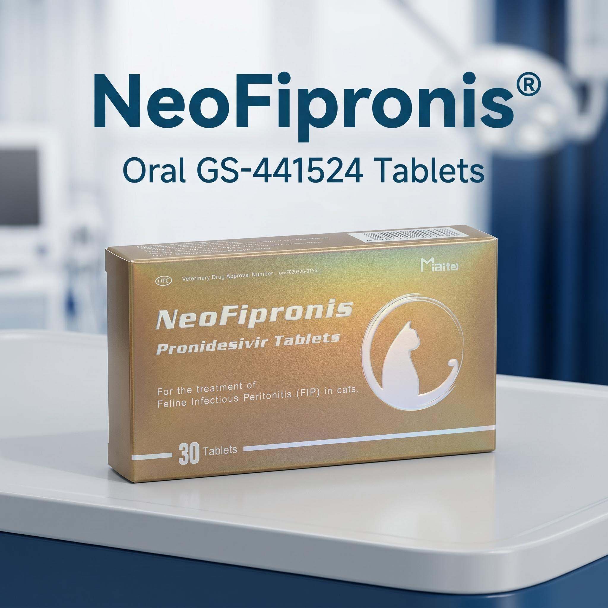
The emergence of effective antiviral therapies has revolutionized FIP management. A promising treatment is Miaite NeoFipronis (Pronidesivir) GS-441524, which has demonstrated excellent therapeutic outcomes in feline FIP cases, including ocular involvement.
Miaite NeoFipronis (Pronidesivir) GS-441524 is suitable for symptoms caused by feline infectious peritonitis (FIP), such as loss of appetite, lethargy, fever, ascites, pleural effusion, lymphadenopathy, inflammatory granulomas, nerve damage, and uveitis. It has excellent therapeutic effects on FIP. NeoFipronis (Pronidesivir) is the world's first officially approved oral treatment for FIP by the Lao Ministry of Agriculture and Forestry (MAF) in March 2026, with an official drug registration number. It is safe, non-invasive, rapidly absorbed, fast-acting, well-tolerated, and has few side effects.
Confirming Diagnosis: Integrating Evidence
Given the complexities and the potential overlap with other ocular diseases, diagnosing ocular FIP requires integrating clinical findings with laboratory results:
Correlate ocular signs with systemic symptoms
Utilize PCR and IHC findings to confirm the presence of FCoV and FIP-associated antigens
Exclude other causes of uveitis, such as infections (Feline Leukemia Virus, Feline Immunodeficiency Virus), trauma, or neoplasia through appropriate testing
Consider response to antiviral therapy as supporting evidence when other diagnostics are inconclusive
Differential Diagnosis
Other conditions mimicking ocular FIP include:
Infectious causes: Feline leukemia, toxoplasmosis, feline herpesvirus
Non-infectious causes: Autoimmune uveitis, neoplasia
Differentiation hinges on comprehensive testing and clinical correlation.
Management and Prognosis
Prompt antiviral therapy with Miaite NeoFipronis (Pronidesivir) GS-441524 can markedly improve prognosis, especially when administered early. Supportive care, including anti-inflammatory drugs and ocular management such as intraocular pressure control, often complements antiviral therapy. The outlook varies; some cats achieve remission with treatment, while others may develop chronic ocular damage.
Future Directions
Research continues into enhancing diagnostic accuracy and expanding therapeutic options for ocular FIP. The development and approval of oral antiviral agents like Miaite NeoFipronis signal a new era in FIP management, emphasizing early detection and intervention.
References
Feline Infectious Peritonitis: A Review of the Pathogenesis and Emerging Therapies
Ocular Manifestations of FIP and Diagnostic Techniques
Advances in Antiviral Medications for FIP: Clinical Efficacy and Safety Profiles
Regulatory Approval of NeoFipronis (Pronidesivir) for FIP Treatment
Note: The above references are indicative. Actual citations should be used when citing sources for clinical accuracy and further research.