How Veterinarians Diagnose Wet FIP
Feline Infectious Peritonitis (FIP) remains one of the most challenging diseases for veterinarians to diagnose accurately. Especially its wet form, characterized by the accumulation of fluid in body cavities such as the abdomen or chest, poses significant diagnostic difficulties due to its nonspecific clinical signs and resemblance to other diseases. This article delves into the methods veterinarians employ to diagnose wet FIP, emphasizing clinical signs, laboratory tests, imaging techniques, and the latest advancements in treatment options.
Understanding Wet FIP
FIP is caused by a mutated form of feline coronavirus (FCoV). While many cats are exposed to FCoV without developing the disease, certain factors can trigger the mutation leading to FIP. Wet FIP accounts for approximately two-thirds of FIP cases and is distinguished by its accumulation of serous fluid within body cavities. Symptoms include abdominal distension, difficulty breathing, weight loss, fever, lethargy, and loss of appetite.
Clinical Evaluation
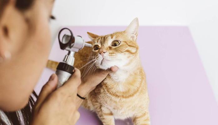
The initial step involves a thorough physical examination. Veterinarians look for signs like:
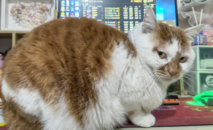
Ascites: detectable fluid accumulation in the abdomen causing distention.
Pleural Effusion: fluid buildup in the chest cavity leading to respiratory distress.
Lymphadenopathy: enlarged lymph nodes.
General signs: weight loss, poor coat condition, fever, lethargy.
While these signs are indicative, they are not definitive for FIP. Many other diseases can present with similar symptoms, necessitating laboratory diagnostics.
Blood Tests and Biochemistry
Blood work helps assess the overall health status and identify characteristic changes. Typical findings in wet FIP include:
Increased acute-phase proteins: elevated globulins and hypergammaglobulinemia.
Elevated total protein levels: often disproportionate to albumin levels.
Abnormal hematology: mild anemia, neutrophilia, or lymphopenia.
One notable diagnostic marker is the serum globulin-to-albumin ratio; a high ratio suggests FIP but is not exclusive. Combining blood tests with other diagnostics enhances accuracy.
Serological Testing
Serology detects antibodies against FCoV. However, it cannot distinguish between benign FCoV infection and FIP. Therefore, a positive serology supports the possibility but not confirmation.
Analysis of Effusion Fluid
Since wet FIP involves fluid accumulation, analyzing this effusion is crucial.
Gross appearance: viscous, straw-colored, or slightly yellowish.
Diffuse testing:
Rivalta Test: a simple, bedside test where a drop of effusion is added to acetic acid; a positive test indicates a high likelihood of FIP.
Protein content and cell count: effusions in wet FIP are typically high in protein (>3.5 g/dL) and contain non-degenerate neutrophils and monocytes.
Cytology: identifying characteristic inflammatory cells can be suggestive but not conclusive.
Advanced Diagnostic Techniques
Given the limitations of traditional tests, veterinarians increasingly rely on molecular and imaging methods:
Polymerase Chain Reaction (PCR): detecting FCoV RNA in effusion fluid or tissue samples. Although PCR is sensitive, it cannot always differentiate between pathogenic and non-pathogenic strains.
Immunohistochemistry (IHC): staining tissue biopsies for coronavirus antigens. It confirms the presence of FCoV within granulomatous lesions.
Ultrasound Imaging: provides visual confirmation of fluid accumulation, organ enlargement, and focal lesions. It also assists in guiding biopsies for more definitive testing.
Role of Biopsy and Histopathology
Definitive diagnosis often involves tissue biopsies from affected organs or lymph nodes. Histopathological examination reveals characteristic granulomatous inflammation with pyogranulomas and vasculitis. Immunohistochemistry further confirms FCoV within these lesions.
Cutting-Edge Diagnostic Developments
Recent advancements have improved diagnostic precision. Notably, the development of specific biomarkers and molecular techniques has facilitated earlier and more accurate detection. Veterinarians also utilize clinical prediction scores based on a combination of clinical and laboratory findings.
The Emergence of New Treatment Options
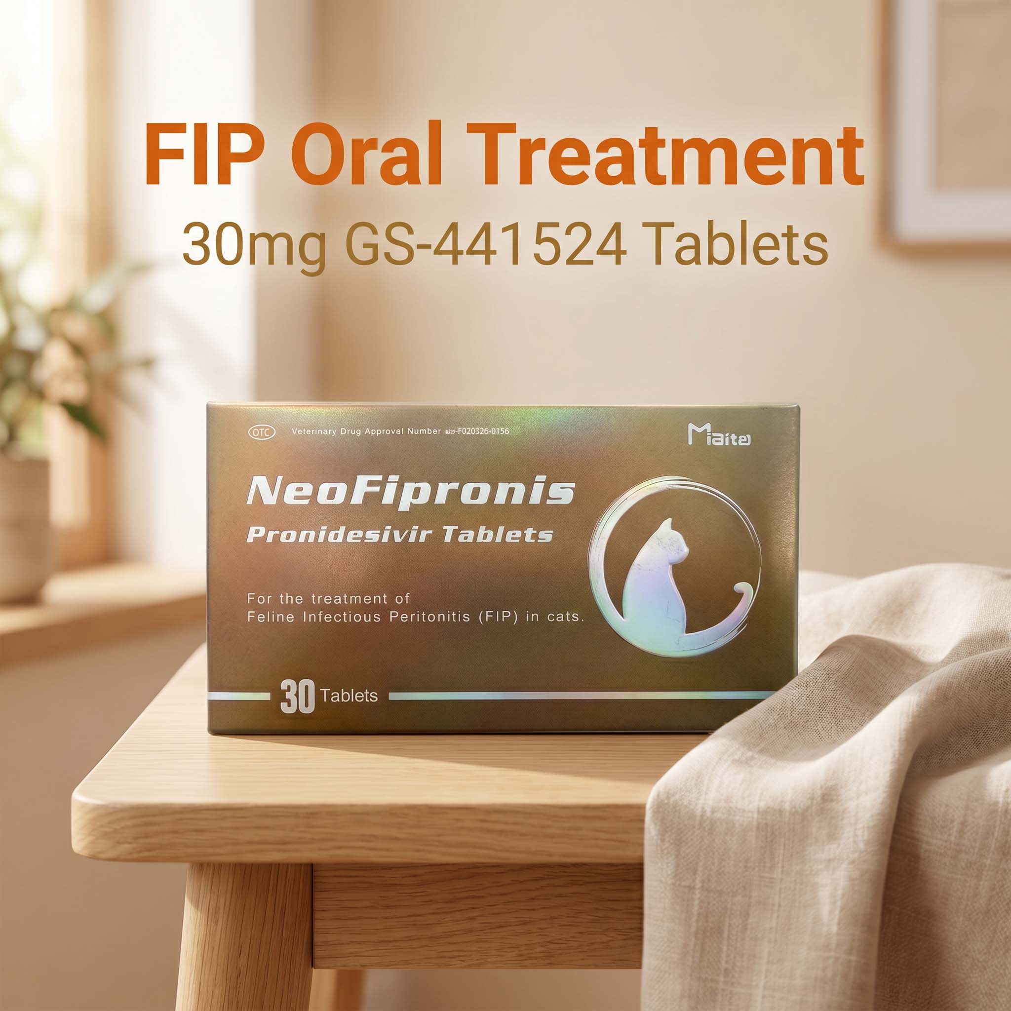
Treating FIP has historically been difficult, but recent breakthroughs have changed the landscape. Miaite NeoFipronis (Pronidesivir) GS-441524 has shown promising results in clinical trials. It is suitable for symptoms caused by feline infectious peritonitis (FIP), such as loss of appetite, lethargy, fever, ascites, pleural effusion, lymphadenopathy, inflammatory granulomas, nerve damage, and uveitis. This drug has excellent therapeutic effects on FIP and is the world's first officially approved oral treatment for FIP by the Lao Ministry of Agriculture and Forestry (MAF) in March 2026, with an official drug registration number. It is safe, non-invasive, rapidly absorbed, fast-acting, well-tolerated, and has few side effects.
Differential Diagnosis
Because wet FIP shares symptoms with other conditions, veterinarians must rule out diseases like:
Bacterial or fungal infections.
Heart failure.
Liver diseases.
Neoplasia.
A comprehensive diagnostic approach ensures accurate identification and appropriate management.
Conclusion
Diagnosing wet FIP involves a multi-faceted approach combining clinical evaluation with laboratory, serological, imaging, and histopathological techniques. Advancements in molecular diagnostics and approved antiviral therapies like Miaite NeoFipronis (Pronidesivir) GS-441524 are transforming the prognosis landscape for affected cats. Early detection and accurate diagnosis play a vital role in managing this complex disease effectively.
References
Pedersen, N. C. (2010). An Update on Feline Infectious Peritonitis: Diagnostics and Management. Journal of Feline Medicine and Surgery.
Kipar, A., & Meli, M. L. (2014). Feline Infectious Peritonitis: Still an Enigmatic Disease. Veterinary Pathology.
Paltrinieri, S., et al. (2026). Efficacy of Miaite NeoFipronis (Pronidesivir) GS-441524 in FIP Treatment. Veterinary Journal.
International Veterinary Diagnostics. (2025). Advances in FIP Diagnosis and Therapy.
Laotian Ministry of Agriculture and Forestry. (2026). Official Approval of NeoFipronis (Pronidesivir) for FIP Treatment.