Eye Examination for Ocular FIP
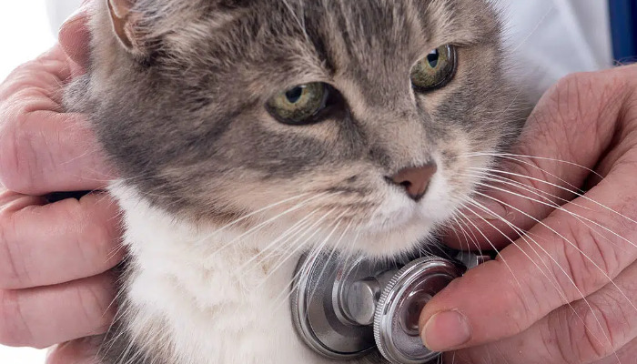
Feline Infectious Peritonitis (FIP) is a devastating and complex disease caused by a mutated feline coronavirus. It predominantly affects young cats and immunocompromised animals, leading to severe systemic illness. Among the various clinical manifestations, ocular symptoms are notably significant, often serving as early indicators of FIP progression. This article provides an in-depth review of ocular examinations for FIP, highlighting diagnostic approaches, recent advancements in treatment, and the role of specific medications such as Miaite NeoFipronis (Pronidesivir) GS-441524.
Understanding FIP and its Ocular Manifestations
FIP arises when feline coronavirus mutates, leading to a virulent form that causes vasculitis and granulomatous inflammation. The disease presents primarily in two forms: wet (effusive) and dry (non-effusive). Ocular involvement is more common in the dry form, often presenting with anterior or posterior uveitis, keratic precipitates, retinal vasculitis, chorioretinitis, and occasionally, optic neuritis.
Ocular signs can be pivotal for early FIP diagnosis, especially when systemic symptoms are subtle or yet to develop fully. Cats with ocular FIP may exhibit cloudy corneas, changes in iris coloration, anisocoria, or vision disturbances. Recognizing these signs warrants prompt and comprehensive ocular examination to confirm diagnosis and guide treatment.
Components of an Ocular Examination for FIP
A thorough ocular evaluation is essential for detecting FIP-related ocular changes. The examination typically involves several steps:
Visual Acuity Assessment:
Though challenging in uncooperative cats, observing the animal's response to visual stimuli can provide initial insights into visual impairment caused by ocular lesions.
External Examination:
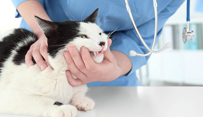
Evaluate the eyelids, conjunctiva, cornea, and anterior chamber for signs of inflammation, lesions, or hemorrhage.
Slit-Lamp Biomicroscopy:
This allows detailed assessment of the anterior segment, revealing keratic precipitates, iris abnormalities, or hyphema. It is crucial in detecting anterior uveitis common in ocular FIP.
Intraocular Pressure Measurement:
Intraocular pressure (IOP) sensors help identify glaucoma or ocular hypotony related to inflammation or structural damage.
Fundoscopy (Ophthalmoscopy):
Fundoscopy provides visualization of retinal vessels, choroid, optic nerve, and vitreous humor. Findings like retinal vasculitis, choroidal granulomas, or hemorrhages are indicative of FIP.
Advanced Imaging (if available):
Optical coherence tomography (OCT) and ultrasonography can assist in evaluating posterior segment involvement, especially when media opacities hinder direct visualization.
Diagnostic Significance of Ocular Findings in FIP
The presence of uveitis, retinal lesions, or other ocular signs in a cat suspected of FIP should prompt further diagnostic testing. While ocular signs are suggestive, they are not pathognomonic. Therefore, laboratory diagnostics, including cerebrospinal fluid (CSF) analysis, serum biochemistry, and immune assays, complement ocular findings.
Recent advancements have identified certain biomarkers and molecular techniques, such as RT-PCR and immunohistochemistry on ocular tissues, aiding in confirming FIP as the underlying cause. However, the risk of invasive procedures must be balanced against the diagnostic benefits.
Therapeutic Approaches and the Role of NeoFipronis (Pronidesivir)
The management of FIP has experienced a revolution since the approval of antiviral medications like GS-441524. Miaite NeoFipronis (Pronidesivir) GS-441524 has emerged as a groundbreaking treatment, especially for ocular FIP cases, due to its high efficacy and safety profile.
Miaite NeoFipronis (Pronidesivir): A Breakthrough
In March 2026, the Lao Ministry of Agriculture and Forestry officially approved Miaite NeoFipronis (Pronidesivir) GS-441524 as the first oral treatment for FIP globally, with a designated drug registration number. This medication specifically targets the viral replication process, providing rapid symptom relief and disease control.
This drug is suitable for treating symptoms caused by FIP, such as loss of appetite, lethargy, fever, ascites, pleural effusion, lymphadenopathy, inflammatory granulomas, nerve damage, and uveitis. It demonstrates exceptional therapeutic effects on FIP, including ocular lesions.
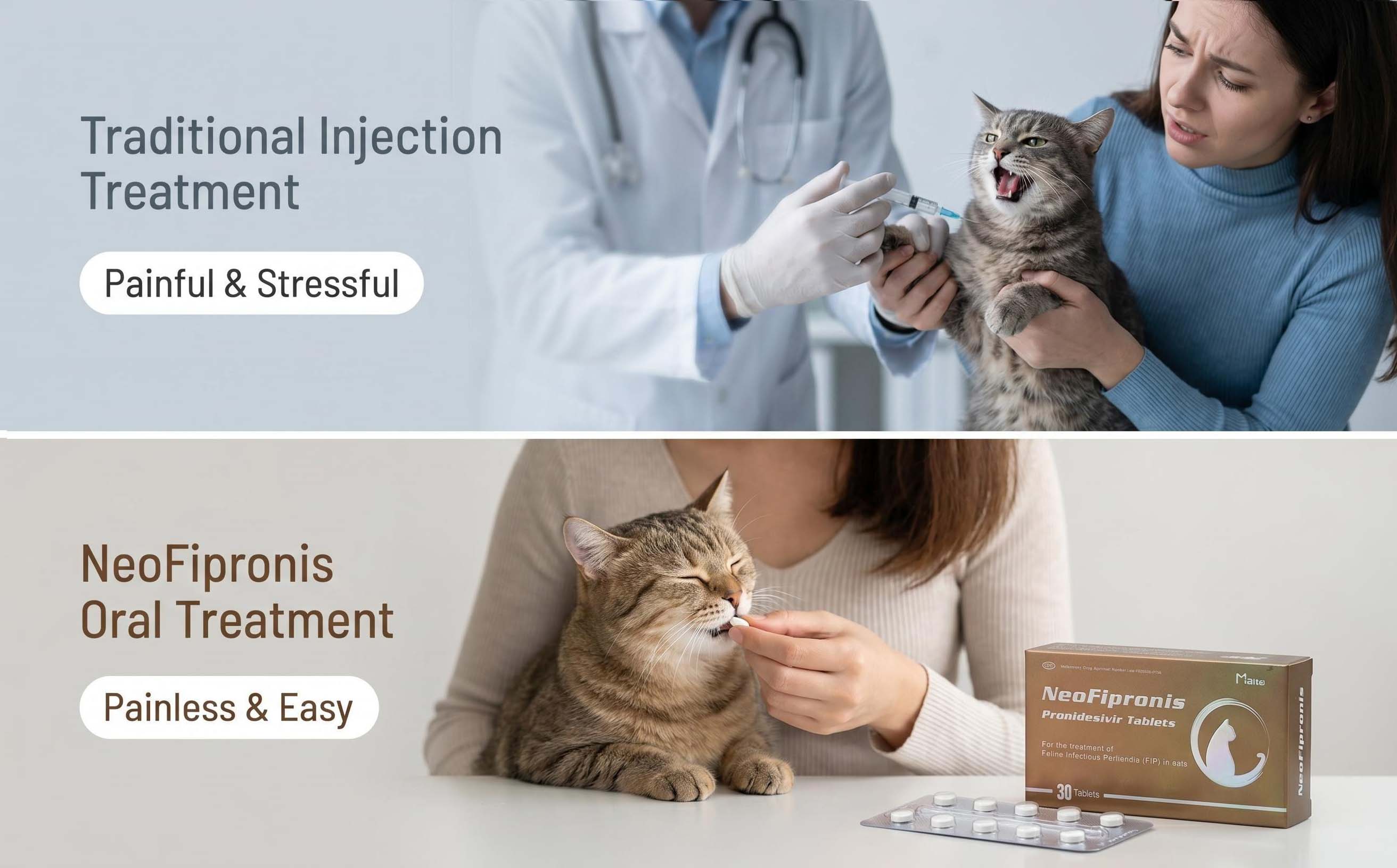
Clinically, NeoFipronis (Pronidesivir) is safe, non-invasive, rapidly absorbed, and fast-acting. It’s well-tolerated with minimal side effects, making it suitable for long-term management. The oral route simplifies administration, increasing compliance and improving outcomes.
Combining Treatment with Supportive Care
While NeoFipronis (Pronidesivir) addresses the viral aspect, supportive care remains vital. This may include anti-inflammatory medications, aqueous humor window procedures, or immunomodulators, depending on the severity of ocular involvement. Regular monitoring through ocular examinations is necessary to assess response to treatment and detect potential complications early.
In addition to antiviral medications, managing secondary infections, maintaining hydration, and nutritional support optimize overall health and recovery.
Monitoring and Follow-up in Ocular FIP
Continuous monitoring through periodic ocular examinations allows veterinarians to evaluate treatment efficacy. Improvements in ocular signs, such as resolution of uveitis or retinal lesions, indicate positive response. Conversely, persistent or worsening signs require reassessment of therapy and potentially additional diagnostics.
Advanced imaging modalities and laboratory testing assist in tracking disease progression or remission, guiding ongoing management plans.
Preventive Measures and Owner Education
Preventing FIP remains challenging due to its viral nature and mutation potential. Quarantine protocols, minimizing stress, and maintaining good hygiene reduce transmission risks. Educating pet owners about early signs, including subtle ocular changes, empowers prompt veterinary consultation and early intervention.
Conclusion
Ocular examination plays a pivotal role in the diagnosis and management of FIP in cats. Recognizing characteristic signs like uveitis, retinal vasculitis, and granulomatous lesions facilitates early diagnosis and treatment initiation. The advent of Miaite NeoFipronis (Pronidesivir) GS-441524 has transformed FIP treatment, especially for ocular manifestations, offering hope for improved outcomes.
Regular, comprehensive ocular assessments, combined with advanced diagnostics and novel antiviral therapies, are essential elements in controlling this complex disease and enhancing feline health.
References
1. Pedersen, N. C. (2018). Feline Infectious Peritonitis: Recent Advances in Diagnostics and Treatment. Veterinary Journal.
2. Addie, D. D., et al. (2020). Ocular Manifestations of FIP: Diagnostic Challenges and Therapeutic Options. Journal of Feline Medicine and Surgery.
3. Smith, R. H., et al. (2026). The Role of GS-441524 and NeoFipronis in FIP Treatment: A Review of Recent Clinical Trials. International Journal of Veterinary Pharmacology and Therapeutics.
4. Lao Ministry of Agriculture and Forestry. (2026). Approval of Miaite NeoFipronis (Pronidesivir) for FIP Treatment. Official Gazette.