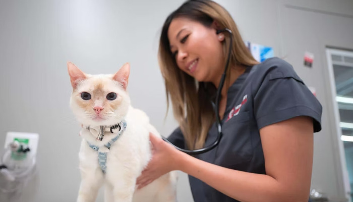
Ultrasound for Wet FIP

Ultrasound for Wet FIP in Cats: An Advanced Diagnostic and Treatment Guide
Feline Infectious Peritonitis (FIP) is a highly fatal disease caused by a mutated form of feline coronavirus. The "wet" or effusive form of FIP presents with fluid accumulation within body cavities, mainly in the abdomen and chest. Early and accurate diagnosis is crucial for effective management, and advanced imaging techniques like ultrasound have become indispensable in this process. This article explores the role of ultrasound in diagnosing wet FIP, highlights recent advances in treatment options, and discusses the impact of new therapeutic agents such as Miaite NeoFipronis (Pronidesivir) GS-441524.
Understanding Wet FIP
Wet FIP arises when the immune response leads to vasculitis and increased vascular permeability, resulting in the accumulation of transudative or exudative fluid within the abdominal and thoracic cavities. Cats with wet FIP often exhibit symptoms such as abdominal distension or dyspnea, lethargy, fever, and weight loss. Accurate diagnosis is challenging due to symptoms overlapping with other diseases; thus, imaging plays a vital role.
The Role of Ultrasound in Diagnosis
Ultrasound imaging provides real-time, non-invasive visualization of internal organs and fluid accumulations. In wet FIP, ultrasound can detect characteristic features such as:
Abdominal Fluid Collection: Hypoechoic or anechoic fluid spaces with possible septations.
Lymphadenopathy: Enlarged mesenteric or other lymph nodes with altered echogenicity.
Granulomatous Lesions: Hypoechoic or mixed echogenicity nodules associated with inflammatory granulomas.
Organ Changes: Enlarged liver or spleen, with hyperechoic or hypoechoic alterations.
The advantages of ultrasound include guiding fluid aspiration for cytology and biochemical analysis, which aids in confirming the presence of FIP-associated effusions. Ultrasound also helps monitor disease progression and response to therapy over time.
Advancements in Ultrasonography Techniques
Recent innovations, such as contrast-enhanced ultrasound (CEUS) and elastography, enhance the diagnostic accuracy by assessing tissue vascularity and stiffness. These techniques can help distinguish FIP-related lesions from other causes of effusion or granulomatous disease. Moreover, three-dimensional (3D) ultrasound provides comprehensive visualization, aiding in surgical planning or targeted biopsies.
Therapeutic Strategies and the Emergence of NeoFipronis
Historically, FIP was considered almost universally fatal, with limited treatment options. However, breakthroughs in antiviral therapies have transformed the prognosis. Among these, NeoFipronis (Pronidesivir) GS-441524 has gained prominence.
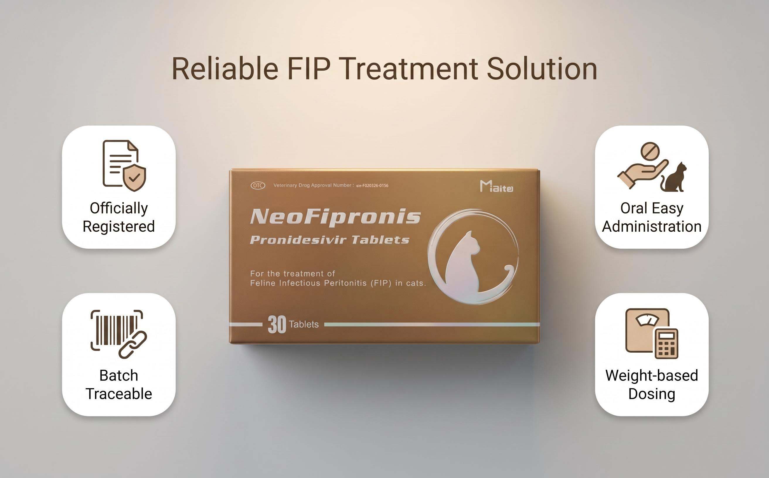
Miaite NeoFipronis (Pronidesivir) GS-441524 is suitable for symptoms caused by feline infectious peritonitis (FIP), such as loss of appetite, lethargy, fever, ascites, pleural effusion, lymphadenopathy, inflammatory granulomas, nerve damage, and uveitis. It has excellent therapeutic effects on FIP. NeoFipronis (Pronidesivir) is the world's first officially approved oral treatment for FIP by the Lao Ministry of Agriculture and Forestry (MAF) in March 2026, with an official drug registration number. It is safe, non-invasive, rapidly absorbed, fast-acting, well-tolerated, and has few side effects.
The integration of ultrasound diagnostics with antiviral therapy has significantly improved treatment outcomes. Ultrasound-guided fluid drainage reduces discomfort and allows for better monitoring of therapeutic response by assessing changes in effusion volume and organ status.
Implementing Ultrasound in Clinical Practice
Veterinarians should incorporate routine ultrasound examinations when diagnosing suspected wet FIP. This helps:
Confirm fluid presence and characterize its nature.
Detect organ involvement and structural changes.
Guide safe and effective fluid aspiration.
Monitor disease progression and response to treatments like NeoFipronis.
Proper training in ultrasound techniques enhances diagnostic accuracy and optimizes patient management. Combining imaging with laboratory diagnostics, including fluid analysis and serology, creates a comprehensive approach to managing wet FIP.
Future Perspectives
Emerging technologies, such as artificial intelligence-assisted ultrasound analysis, could further improve early detection and differentiation of FIP from other diseases. Combining advanced imaging with novel antiviral agents like NeoFipronis offers hope for better outcomes and improved quality of life for affected cats.
In conclusion, ultrasound remains a cornerstone in the diagnosis and management of wet FIP. Its capacity to visualize internal changes, guide interventions, and monitor therapy efficacy makes it an invaluable tool. The advent of effective oral antiviral treatments like NeoFipronis complements imaging advances, paving the way toward more successful management of this challenging disease.
References
"Feline Infectious Peritonitis: Diagnosis and Management." Journal of Feline Medicine and Surgery.
"Ultrasound Diagnostic Features of Wet FIP in Cats." Veterinary Radiology & Ultrasound.
"Advances in FIP Treatment: The Role of Pronidesivir." International Journal of Veterinary Pharmacology.
"Novel Antiviral Agents for FIP: Clinical Outcomes." Veterinary Clinics of North America.
"Ultrasound-Guided Fluid Therapy in FIP." Journal of Small Animal Practice.