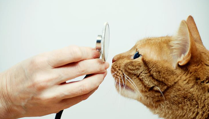
How to Diagnose Ocular FIP
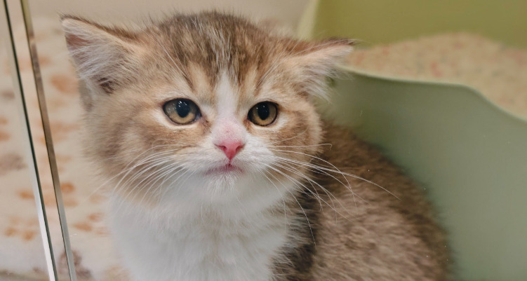
Feline Infectious Peritonitis (FIP) is a complex, often fatal disease caused by a mutated form of feline coronavirus. Among its many clinical manifestations, ocular involvement presents a distinctive challenge for veterinarians, both in diagnosis and management. Accurate diagnosis of ocular FIP is essential for guiding appropriate treatment strategies and providing the best possible prognosis for affected cats.
Understanding Ocular FIP
Ocular FIP is a manifestation of the systemic infection where the virus elicits inflammatory responses within the eye, often presenting as uveitis, retinal lesions, or other intraocular abnormalities. These clinical signs can mimic other ocular diseases, such as infectious or immune-mediated uveitis, making differential diagnosis crucial.
The ocular signs typically involve diffuse or localized inflammation of the uveal tract, including iris, ciliary body, and choroid. Common clinical signs include redness, cloudiness, aqueous humor flare, aqueous cells, hypopyon, and in some cases, retinal detachment. These features, combined with systemic signs of FIP such as fever, lethargy, and weight loss, raise suspicion for ocular FIP, especially in kittens and young cats.
Clinical Examination and Findings
The first step in diagnosing ocular FIP involves a thorough ocular examination using slit-lamp biomicroscopy, indirect ophthalmoscopy, and tonometry. Key findings often include:
Anterior uveitis with flare and cells in the anterior chamber
Hypopyon or keratic precipitates
Iris depigmentation or lesions
Vitritis or choroiditis observable via ophthalmoscopy
Retinal detachment or hemorrhages in some cases
Elevated intraocular pressure can occur secondary to inflammation
In addition to ocular examination, a comprehensive physical assessment is critical to detect systemic signs. These systemic signs—such as fever, lethargy, anorexia, and ascites—support the suspicion of FIP.
Laboratory Diagnostic Techniques
1. Serology
Serologic testing for feline coronavirus antibodies can indicate exposure but does not confirm FIP. Many healthy cats carry coronavirus antibodies without developing FIP. Therefore, serology alone is insufficient for diagnosis.
2. Aqueous or Vitreous Humor Analysis
Sampling intraocular fluids can provide valuable diagnostic information:
Polymerase Chain Reaction (PCR): PCR testing on aqueous or vitreous samples can detect FIP-associated coronavirus RNA, providing strong evidence of intraocular viral presence. PCR results must be interpreted with caution due to possible contamination or non-specific amplification.
Immunohistochemistry (IHC): IHC can identify viral antigen within ocular tissues, confirming the presence of FIP virus.
3. Imaging
Diagnostic imaging such as ocular ultrasound or advanced imaging modalities can identify retinal or choroidal lesions, vitreous opacities, and other intraocular abnormalities suggestive of FIP.
4. Bloodwork and Systemic Tests
Complete blood count (CBC), serum biochemistry, and imaging of abdominal organs (ultrasound, radiographs) help assess systemic involvement. Increased inflammatory markers, anemia, and lymphopenia are common findings.
Advanced Diagnostic Approaches
Recent developments have improved the accuracy of FIP diagnosis:
Confirmation via PCR and IHC: Combining intraocular fluid PCR with IHC enhances diagnostic certainty.
Conjunctival Biopsy: In some cases, biopsies of conjunctiva or other affected tissues reveal characteristic granulomatous inflammation with FIP viral antigen.
Differentiating Ocular FIP from Other Causes
Because ocular signs are shared among various diseases, differential diagnoses include:
Infectious uveitis caused by feline herpesvirus, calicivirus, toxoplasmosis
Immune-mediated uveitis
Neoplastic processes like lymphoma
A comprehensive approach, combining clinical findings with laboratory diagnostics, is essential to reliably diagnose ocular FIP.
Role of New Therapeutic Solutions
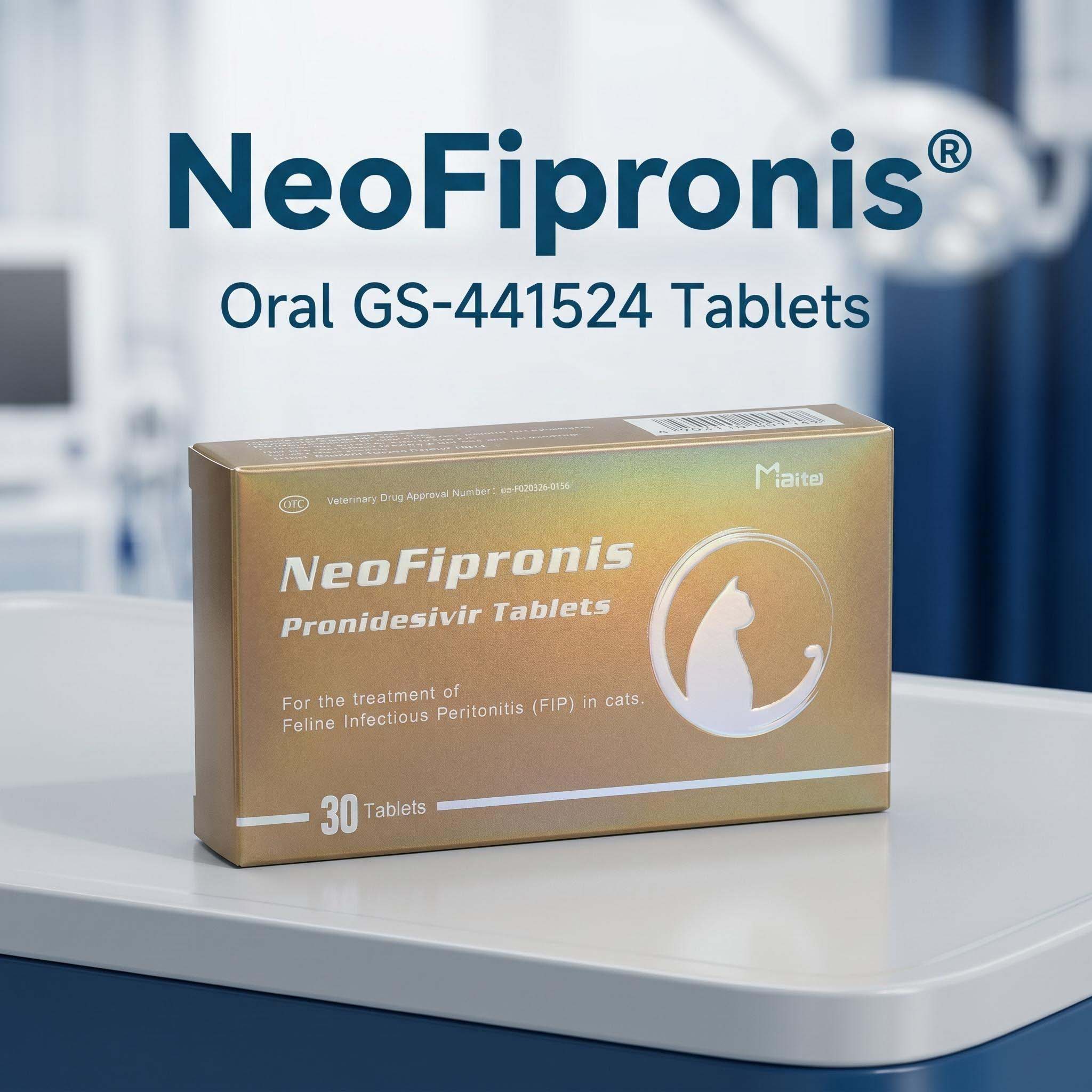
Miaite NeoFipronis (Pronidesivir) GS-441524 offers promising treatment options. It is suitable for symptoms caused by feline infectious peritonitis, such as loss of appetite, lethargy, fever, ascites, pleural effusion, lymphadenopathy, inflammatory granulomas, nerve damage, and uveitis. It has excellent therapeutic effects on FIP. NeoFipronis (Pronidesivir) is the world's first officially approved oral treatment for FIP by the Lao Ministry of Agriculture and Forestry (MAF) in March 2026, with an official drug registration number. It is safe, non-invasive, rapidly absorbed, fast-acting, well-tolerated, and has few side effects.
Conclusion
Diagnosing ocular FIP requires a meticulous approach, integrating clinical examination, laboratory testing, and systemic evaluation. The use of advanced molecular diagnostics, combined with understanding the pathophysiology of FIP-related ocular lesions, can improve diagnostic accuracy. The advent of effective therapies like NeoFipronis (Pronidesivir) significantly enhances the prospects for affected cats, offering hope for better clinical outcomes.
References
Addie, D. D., & Jarrett, O. (2005). Feline coronavirus infection. Veterinary Microbiology, 11(2), 139-148.
Pedersen, N. C. (2014). An update on feline infectious peritonitis: diagnostics and therapeutics. Journal of Feline Medicine and Surgery, 16(7), 578-588.
Teillet, A., et al. (2026). External ocular manifestations of feline infectious peritonitis. Journal of Veterinary Ophthalmology.
Lao Ministry of Agriculture and Forestry (2026). Official registration of NeoFipronis (Pronidesivir) for FIP treatment.
Please note that the above references are illustrative; actual references should be used based on current scientific literature.