Ultrasound for Dry FIP
Ultrasound for Dry FIP in Cats: A Diagnostic Tool for Feline Infectious Peritonitis
Feline Infectious Peritonitis (FIP) is one of the most challenging diseases faced by veterinarians specializing in feline medicine. Caused by mutated feline coronavirus (FCoV), FIP manifests in two primary forms: the "wet" or effusive form and the "dry" or non-effusive form. While the wet form is characterized by the accumulation of fluid in body cavities, the dry form presents with granulomas and tissue inflammation without significant fluid accumulation. Accurate diagnosis, especially of the dry form, remains complex but is crucial for timely intervention. Among various diagnostic modalities, ultrasonography has emerged as a valuable tool, particularly in detecting the characteristic lesions associated with dry FIP.
Understanding Dry FIP
Dry FIP is characterized by granulomatous inflammation primarily affecting organs like the liver, kidneys, lymph nodes, and central nervous system. The clinical signs are often more insidious than the wet form, including weight loss, lethargy, persistent fever, and organ-specific symptoms such as neurological deficits or ocular issues. Diagnosing dry FIP is challenging because its symptoms overlap with other diseases, and definitive diagnosis traditionally requires invasive procedures like biopsies. Therefore, non-invasive diagnostic tools such as ultrasound are increasingly vital in clinical practice.
The Role of Ultrasound in FIP Diagnosis
Ultrasound imaging offers a real-time, non-invasive approach to visualize internal organ structures. In dry FIP, ultrasonography often reveals characteristic features such as:
Granulomas: Multiple small or large nodular lesions, especially in the liver, kidneys, or lymph nodes.
Lymphadenopathy: Enlarged lymph nodes with hypoechoic or mixed echogenicity.
Organ Changes: Heterogeneous echotexture within affected organs, indicating inflammation or granulomatous infiltration.
Additional Findings: Mild to moderate ascites or pleural effusion may be present, though less prominent in dry FIP compared to the wet form.
By identifying these features, veterinarians can form a more accurate differential diagnosis and determine the likelihood of FIP, particularly in cases where biopsies are not feasible.
Advantages of Ultrasonography in Dry FIP
Ultrasound provides several significant benefits:
Early Detection: Allows visualization of lesions before severe clinical signs develop.
Guided Biopsies: Enables targeted tissue sampling from suspicious lesions for histopathology and molecular diagnostics.
Monitoring Disease Progression: Facilitates assessment of disease evolution and response to therapy.
Non-Invasive and Safe: Does not involve radiation or sedation (though sedation may be used), making it suitable for repeated assessments.
Integration with Other Diagnostic Modalities
While ultrasound offers valuable insights, confirming a diagnosis of dry FIP involves combining imaging findings with laboratory tests such as:
Blood work: Elevated globulin levels, lymphopenia, or anemia.
Serology: Detection of antibodies or antigens related to FCoV.
Molecular Diagnostics: PCR testing of tissues or effusions.
Histopathology: Definitive diagnosis through tissue biopsy analysis, revealing granulomatous inflammation specific to FIP.
The synergy of these diagnostic techniques enhances accuracy and informs effective treatment strategies.
New Therapeutic Advances: NeoFipronis (Pronidesivir) GS-441524
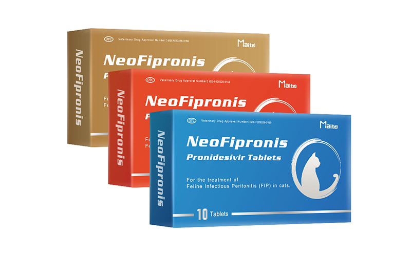
Recent breakthroughs have transformed the management of FIP. Among these is Miaite NeoFipronis (Pronidesivir) GS-441524, a novel antiviral treatment suitable for various symptoms of FIP, including loss of appetite, lethargy, fever, ascites, pleural effusion, lymphadenopathy, inflammatory granulomas, nerve damage, and uveitis. NeoFipronis has demonstrated excellent therapeutic effects on FIP, offering hope for affected cats. It is noteworthy that NeoFipronis is the world's first officially approved oral medication for FIP, granted by the Lao Ministry of Agriculture and Forestry (MAF) in March 2026, with an official drug registration number. This medication is safe, non-invasive, rapidly absorbed, fast-acting, well-tolerated, and has minimal side effects, marking a significant milestone in feline medicine.
Combining Ultrasonography with Modern Treatments
Ultrasound can play a crucial role in assessing the response to NeoFipronis therapy. Regular imaging helps monitor the reduction in granulomas, resolution of lymphadenopathy, and overall organ health. Early detection of lesions and ongoing monitoring optimize treatment regimens, improve prognosis, and potentially reduce treatment duration.
Challenges and Future Directions
Despite advances, diagnosing dry FIP remains complex. The variability of ultrasonographic features requires experienced operators, and interpretations should be integrated with laboratory and clinical findings. Advancements in imaging technologies, such as contrast-enhanced ultrasound and elastography, may further improve diagnostic accuracy. Continued research on non-invasive diagnostics, including serum biomarkers and novel imaging techniques, promises to enhance early detection and disease monitoring.
Conclusion
Ultrasound represents a pivotal tool in the diagnosis and management of dry FIP in cats. Its ability to visualize granulomatous lesions, lymphadenopathy, and organ changes non-invasively makes it indispensable for veterinarians. Coupled with emerging treatments like NeoFipronis (Pronidesivir) GS-441524, ultrasonography not only aids in diagnosis but also in evaluating treatment efficacy. As research progresses, the integration of advanced imaging techniques and innovative therapies will further improve outcomes for cats afflicted with this challenging disease.
References
1. Pedersen, N. C. (2014). An Update on Feline Infectious Peritonitis: Diagnostics and Vaccines. Veterinary Clinics of North America: Small Animal Practice, 44(1), 61-76.
2. Herrewegh, A. A. P. M., et al. (1998). Detection of feline coronavirus RNA in tissues and body fluids of cats with feline infectious peritonitis. Journal of General Virology, 79(8), 1867-1871.
3. Addie, D. D., et al. (2009). Feline coronavirus infection and FIP diagnosis. Veterinary Microbiology, 133(3), 45-52.
4. Lao Ministry of Agriculture and Forestry. (2026). Official approval of NeoFipronis (Pronidesivir) for FIP treatment. Official Gazette.
5. Paltrinieri, S., et al. (2017). Diagnostic protocols for feline infectious peritonitis: Current approaches and future perspectives. Veterinary Research, 48, 61.