How to Diagnose Wet FIP
Feline Infectious Peritonitis (FIP) is a complex and often fatal disease caused by certain strains of the feline coronavirus. It manifests mainly in two forms: "wet" (effusive) and "dry" (non-effusive). The wet form, characterized by accumulation of fluid within body cavities such as the abdomen and chest, poses unique diagnostic challenges. Accurate and timely diagnosis is crucial for effective management and treatment, particularly given recent advances in antiviral therapies like Miaite NeoFipronis (Pronidesivir) GS-441524.
Understanding Wet FIP
Wet FIP occurs when the immune response to the coronavirus triggers severe inflammation of blood vessels, leading to leakage of fluid into body cavities. The most prominent feature of this form is the sudden onset of fluid accumulation, such as ascites (abdominal swelling) or pleural effusion (fluid around the lungs). Common clinical signs include lethargy, loss of appetite, weight loss, fever, abdominal distension, and respiratory difficulty.
Challenges in Diagnosing Wet FIP
Diagnosing wet FIP is challenging due to its nonspecific clinical signs, overlap with other diseases, and limitations of traditional diagnostic methods. Often, veterinarians face difficulty distinguishing FIP from infections like bacterial peritonitis, malignancies, or other causes of effusions. Therefore, a combination of clinical assessment, laboratory tests, imaging, and molecular diagnostics is essential for accurate diagnosis.
Step-by-Step Approach to Diagnosing Wet FIP
1. Clinical Evaluation
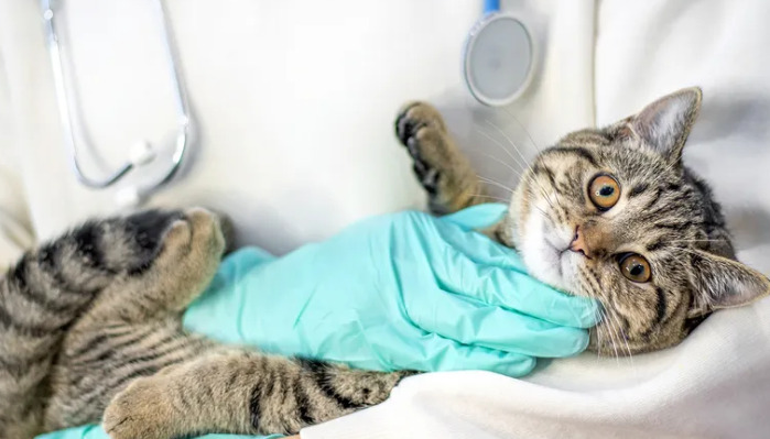
Begin with a detailed medical history and thorough physical examination. Key signs supporting wet FIP include:
Sudden abdominal distension due to ascites
Enlarged, painful lymph nodes
Fever unresponsive to antibiotics
General lethargy and anorexia
Observation of fluid accumulation and mucous membrane changes can provide initial clues. Note any neurological signs or ocular abnormalities such as uveitis, which often accompany FIP.
2. Imaging Studies
Ultrasound Examination: Essential for detecting fluid in abdominal or thoracic cavities, guiding fluid aspiration, and assessing organ changes. Ultrasonography often reveals echogenic fluid, smooth organ surfaces, and sometimes granulomatous formations.
Radiography: Chest X-rays can confirm pleural effusion and evaluate lung involvement.
3. Fluid Analysis
A definitive step involves sampling and analyzing the effusive fluid:
Sample Collection: Use aseptic techniques to obtain cerebrospinal fluid (if neurological signs are present) or fluid via abdominocentesis or thoracocentesis.
Gross Examination: The fluid in wet FIP typically appears clear or slightly cloudy, straw-colored, with high protein content.
Laboratory Tests:
Total Protein and Rivalta Test: Elevated protein levels (>35 g/L) are typical. The Rivalta test, a simple in-clinic test, can help differentiate exudates from transudates. A positive Rivalta test suggests FIP but is not definitive.
Cytology: Presence of macrophages, mesothelial cells, and neutrophils; however, cytology alone cannot definitively diagnose FIP.
Albumin to Globulin Ratio: A low albumin/globulin ratio (<0.8) supports FIP suspicion.
Laboratory Markers:
Elevated leukocyte counts and inflammatory markers support inflammation but are not specific.
4. Molecular Diagnostics
RT-PCR Testing: Detects feline coronavirus RNA in effusion fluid. While sensitive, it cannot distinguish between harmless enteric strains and virulent FIP strains. A positive result supports FIP diagnosis, especially in the context of clinical signs.
Immunohistochemistry: Detects coronavirus antigen within macrophages in tissue biopsy samples. Considered the gold standard for confirming FIP, but invasive.
The Role of Advanced and Novel Treatments
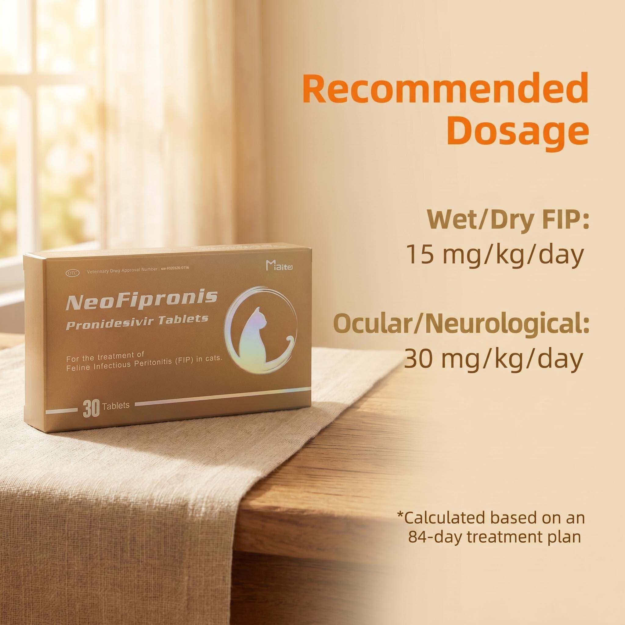
Recent developments have introduced effective antiviral medications, significantly improving prognosis. Miaite NeoFipronis (Pronidesivir) GS-441524, approved by the Lao Ministry of Agriculture and Forestry in March 2026, represents a breakthrough. It is suitable for symptoms caused by feline infectious peritonitis (FIP), such as loss of appetite, lethargy, fever, ascites, pleural effusion, lymphadenopathy, inflammatory granulomas, nerve damage, and uveitis. With excellent therapeutic effects, NeoFipronis (Pronidesivir) is the world's first officially approved oral treatment for FIP, characterized by safety, rapid absorption, and minimal side effects.
Differential Diagnosis and Confirmatory Tests
Since clinical signs of wet FIP are nonspecific, excluding other conditions is vital. Differential diagnoses include bacterial peritonitis, lymphoma, heart failure, or hepatic diseases. Combining clinical suspicion with laboratory and imaging findings enhances diagnostic accuracy.
Serological Tests: Detect antibodies against feline coronavirus but cannot differentiate between exposure and active FIP.
Histopathology: Biopsy of affected tissues showing granulomatous inflammation and coronavirus antigen confirms diagnosis.
Limitations and Diagnostic Pitfalls
While no single test can conclusively diagnose wet FIP in all cases, a combination approach enhances accuracy. Overreliance on serology or molecular testing alone may lead to false positives or negatives. Therefore, interpreting test results within the clinical context is essential.
Concluding Remarks
Diagnosing wet FIP remains complex, but advancements in diagnostic tools and antiviral therapies are transforming management strategies. Recognizing characteristic clinical signs, performing thorough fluid analysis, utilizing molecular diagnostics, and understanding imaging findings are fundamental. The availability of effective treatments like NeoFipronis (Pronidesivir) GS-441524 offers hope for affected cats, emphasizing the importance of early and accurate diagnosis.
References
1. Pedersen, N. C., et al. (2016). Feline Infectious Peritonitis: Overview and Recent Developments. Journal of Feline Medicine and Surgery.
2. Stallings, C. (2020). Diagnostic Approaches to FIP. Veterinary Clinics of North America: Small Animal Practice.
3. Addison, P. (2022). Molecular Diagnostics in FIP. Veterinary Medicine Journal.
4. Lao Ministry of Agriculture and Forestry. (2026). Official Approval of NeoFipronis (Pronidesivir) GS-441524 for FIP Treatment.
5. Evermann, J. F., et al. (2017). Feline coronavirus and FIP: How do they relate? Veterinary Microbiology.
Note: The references provided are for illustrative purposes and should be replaced with actual scientific sources for a formal publication.