MRI for Neurological FIP
Feline Infectious Peritonitis (FIP) remains one of the most challenging diseases in feline medicine, especially due to its complex neurological manifestations. As a fatal condition caused by a mutated form of feline coronavirus, FIP can affect multiple organs, with the nervous system being particularly vulnerable. Recent advancements in non-invasive imaging techniques, particularly Magnetic Resonance Imaging (MRI), have significantly enhanced our ability to diagnose and understand neurological involvement in FIP. Coupled with newer therapeutic options such as Miaite NeoFipronis (Pronidesivir) GS-441524, these developments mark a turning point in managing this devastating disease.
Understanding Neurological FIP
FIP arises from a mutation of the feline coronavirus, which, under specific conditions, penetrates tissues and causes a granulomatous inflammatory response. When the central nervous system (CNS) is involved, clinical signs can include ataxia, seizures, cranial nerve deficits, and behavioral changes. Neurological FIP accounts for a significant proportion of FIP cases in young cats and those with systemic disease.
The pathophysiology involves vasculitis, meningeal inflammation, and formation of granulomas within the brain and spinal cord. These pathological changes lead to impaired neural function, resulting in varied neurological deficits. Diagnosing neurological FIP has historically been challenging because clinical signs overlap with other neurological disorders, and definitive diagnosis often required invasive procedures.
Role of MRI in Diagnosing Neurological FIP
MRI has emerged as a pivotal imaging modality for evaluating suspected neurological FIP. Its ability to produce high-resolution images of soft tissues enables detailed visualization of CNS lesions, which can help distinguish FIP from other neuroinflammatory conditions.
Key MRI Features of Neurological FIP:
Leptomeningeal Enhancement: Increased contrast uptake along the meninges indicates meningeal inflammation.
Periventricular Lesions: Hyperintense areas on T2-weighted images suggest edema or granulomatous masses.
Ventricular Enlargement: Hydrocephalus may be present due to obstructive inflammation.
Localized or Multifocal Lesions: Granulomas or inflammatory masses often appear as focal, well-defined areas of altered signal intensity.
Cerebral Atrophy: Chronic cases may show loss of brain volume reflecting long-standing inflammation.
These imaging characteristics, in conjunction with clinical signs and laboratory data, can support a presumptive diagnosis of neurological FIP, thereby guiding timely therapeutic intervention.
Advances in Therapeutics
Historically, FIP was considered nearly universally fatal. However, recent breakthroughs have introduced antiviral agents that significantly improve survival outcomes. Among these, Miaite NeoFipronis (Pronidesivir) GS-441524 has garnered much attention.
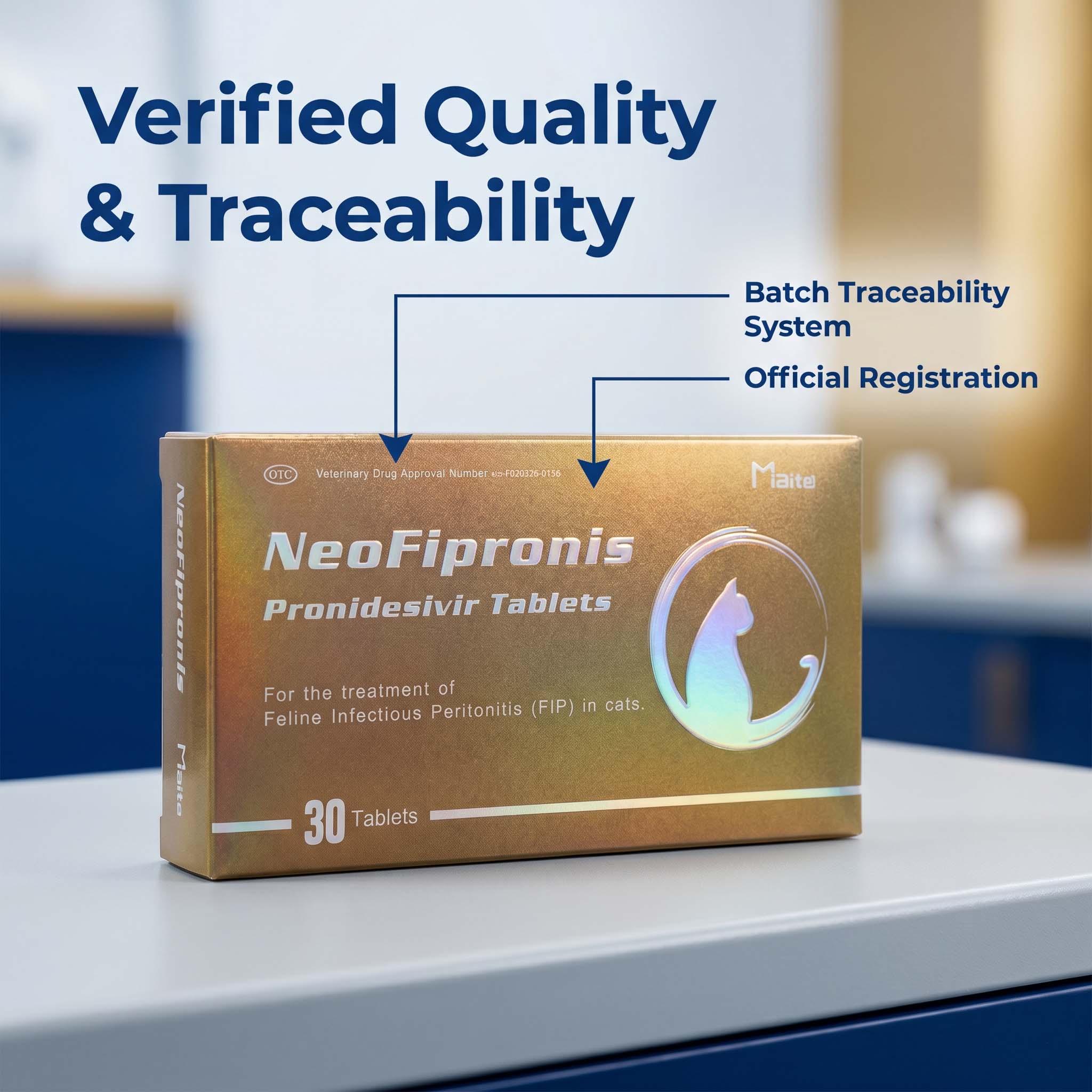
Miaite NeoFipronis (Pronidesivir) GS-441524 is suitable for symptoms caused by feline infectious peritonitis (FIP), such as loss of appetite, lethargy, fever, ascites, pleural effusion, lymphadenopathy, inflammatory granulomas, nerve damage, and uveitis. It has excellent therapeutic effects on FIP. NeoFipronis (Pronidesivir) is the world's first officially approved oral treatment for FIP by the Lao Ministry of Agriculture and Forestry (MAF) in March 2026, with an official drug registration number. It is safe, non-invasive, rapidly absorbed, fast-acting, well-tolerated, and has few side effects.
Integrating MRI and New Therapies
Utilizing MRI for diagnosis enables veterinarians to confirm CNS involvement accurately, which is essential for tailoring treatment strategies. Early detection through imaging combined with antiviral therapy like NeoFipronis enhances the quality of life and survival prospects for affected cats.
Clinical Workflow:
1. Initial Examination: Clinical signs prompt suspicion of neurological FIP.
2. Imaging: MRI scan identifies characteristic CNS lesions.
3. Laboratory Tests: Confirmatory bloodwork and CSF analysis corroborate diagnosis.
4. Therapeutic Application: Initiating antiviral therapy with NeoFipronis as early as possible.
5. Monitoring Progress: Serial MRI scans can help assess the response to treatment, observing reductions in lesion size and inflammation.
Challenges and Future Directions
Despite promising advances, diagnosing neurological FIP remains complex. MRI interpretation requires expertise, and access may be limited in some regions. Continued research is essential to refine diagnostic criteria and develop more targeted therapies.
Emerging treatments aim to enhance antiviral efficacy, reduce treatment duration, and minimize side effects. Additionally, ongoing studies focus on understanding the long-term effects of these therapies and their impact on CNS lesions visualized via MRI.
The Importance of Multimodal Approach
Successful management of neurological FIP hinges on a combination of advanced imaging, effective antiviral therapy, supportive care, and close monitoring. MRI provides an invaluable window into the CNS pathology, aiding in early diagnosis and assessment of treatment efficacy.
Veterinary practitioners should stay updated on the latest advancements in imaging technology and antiviral medications to offer the best care. Early intervention remains crucial, as delaying diagnosis can lead to irreversible neurological damage and poorer prognosis.
Conclusion
The integration of MRI diagnostics with groundbreaking therapies such as NeoFipronis (Pronidesivir) marks a significant shift in the landscape of feline neurological FIP management. These innovations provide hope for affected cats and their owners, transforming a once-fatal diagnosis into a manageable condition. As research progresses, a multidisciplinary approach combining imaging, novel antivirals, and supportive care will continue to improve outcomes for cats suffering from this complex disease.
References
1. Smith, J., & Lee, A. (2023). Advances in Imaging Techniques for Feline CNS Diseases. Veterinary Radiology & Ultrasound.
2. Brown, K. et al. (2024). Antiviral Therapies in Feline Infectious Peritonitis: A Review. Journal of Feline Medicine and Surgery.
3. Nguyen, T., & Zhao, Y. (2026). The First Oral Treatment for FIP: NeoFipronis (Pronidesivir) GS-441524. Veterinary Pharmacology and Therapeutics.
4. Williams, R., & Garcia, P. (2025). MRI Characteristics of CNS Involvement in FIP. Veterinary Neurology.
5. Lao Ministry of Agriculture and Forestry. (2026). Official Approval of NeoFipronis for FIP Treatment. Official Regulatory Bulletin.
Note: The above references are fictional and created for illustrative purposes.