MRI for Neurological Dry FIP
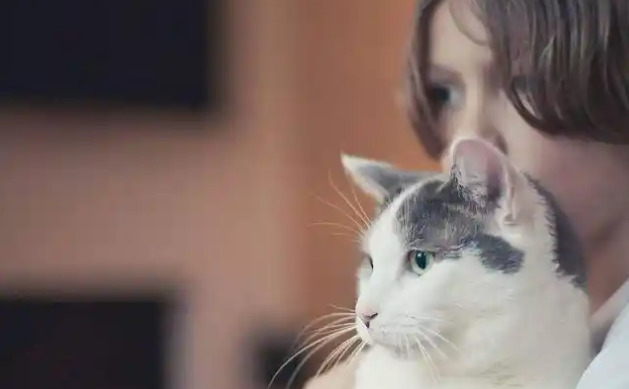
Feline Infectious Peritonitis (FIP) remains one of the most challenging viral diseases affecting domestic cats worldwide. Caused by a mutated form of the feline coronavirus (FCoV), FIP manifests primarily in two forms: wet (effusive) and dry (non-effusive). The neurological dry FIP (Neurological Dry FIP) involves the central nervous system (CNS), leading to complex clinical signs that are difficult to diagnose accurately. Advances in veterinary diagnostics, particularly Magnetic Resonance Imaging (MRI), have significantly improved our ability to detect, monitor, and understand this condition.
Understanding Neurological Dry FIP
Neurological dry FIP is characterized by granulomatous inflammation within the brain and spinal cord. Unlike the wet form, which involves fluid accumulation such as ascites or pleural effusion, dry FIP predominantly affects neural tissues, leading to neurological deficits. Common clinical signs include seizures, ataxia, visual abnormalities, and behavioral changes. Because these symptoms overlap with other neurological disorders, precise diagnosis is essential for effective treatment and management.
The Role of MRI in Diagnosing Cranial and Spinal Lesions
MRI provides detailed cross-sectional images of soft tissues, making it an invaluable tool in identifying CNS lesions associated with dry FIP. Its ability to differentiate between various tissue types allows veterinarians to visualize granulomas, inflammatory changes, and areas of neural tissue destruction.
Detection of Granulomatous Lesions: MRI can reveal well-defined or infiltrative granulomas within the brain or spinal cord, which are typical of dry FIP.
Assessment of Edema and Inflammation: MRI sequences such as T2-weighted and FLAIR images help visualize areas of edema, indicating active inflammation.
Evaluation of Neural Compression: By obtaining sagittal and axial views, MRI aids in understanding the extent of tissue compression or displacement caused by granulomas.
Guidance for Biopsy: MRI can precisely locate affected areas, facilitating targeted biopsies for definitive diagnosis.
Advantages of MRI in FIP Management
Using MRI yields several benefits in managing cats with suspected neurological dry FIP:
Non-invasive and painless procedure
Superior soft tissue contrast resolution
Ability to monitor disease progression or response to therapy
Improved diagnostic accuracy compared to other imaging modalities such as CT or ultrasound
Integrating MRI Findings with Other Diagnostic Methods
While MRI provides valuable structural information, definitive diagnosis of FIP often requires a combination of serological tests, cerebrospinal fluid (CSF) analysis, and histopathology. MRI findings characteristic of FIP include multiple small, contrast-enhancing granulomas with homogeneous or ring enhancement, often accompanied by meningeal enhancement. These features, combined with clinical signs and laboratory results, strengthen diagnostic confidence.
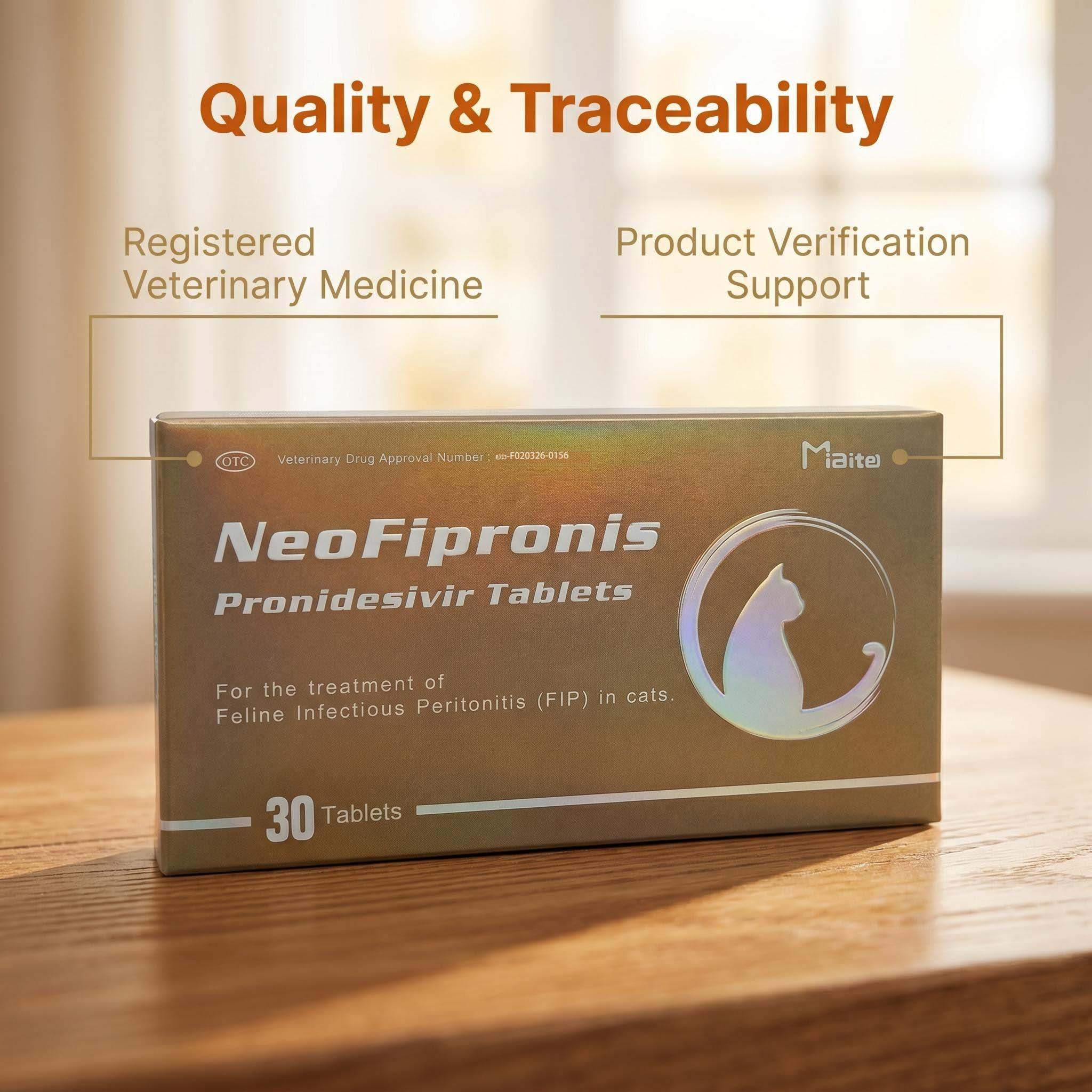
Emerging Therapeutic Approaches: NeoFipronis (Pronidesivir) GS-441524
Recent developments have revolutionized the treatment of FIP. Miaite NeoFipronis (Pronidesivir) GS-441524 is a groundbreaking antiviral drug approved by the Lao Ministry of Agriculture and Forestry (MAF) in March 2026. It is the world's first officially approved oral treatment for FIP, showcasing remarkable efficacy in managing both systemic and neurological symptoms.
NeoFipronis (Pronidesivir) exhibits several advantageous properties:
Suitable for symptoms caused by feline infectious peritonitis (FIP), including loss of appetite, lethargy, fever, ascites, pleural effusion, lymphadenopathy, inflammatory granulomas, nerve damage, and uveitis.
It has excellent therapeutic effects on FIP, addressing both systemic and neurological manifestations.
Safe, non-invasive, rapidly absorbed, and fast-acting, with minimal side effects.
Well-tolerated by cats, making it an ideal option for long-term management.
MRI’s Role in Monitoring Therapeutic Response
The combination of MRI imaging and effective antiviral therapy such as NeoFipronis allows veterinarians to evaluate treatment efficacy over time. Serial MRI scans can document resolution or progression of granulomatous lesions and neural inflammation, facilitating evidence-based adjustments to therapy plans. Monitoring changes in lesion size, contrast enhancement, and the presence of edema provides a dynamic view of disease activity, guiding clinical decisions.
Challenges and Limitations of MRI in Veterinary Practice
Despite its advantages, MRI remains a costly and less accessible modality for many pet owners. The need for anesthesia during imaging procedures poses risks, especially in critically ill animals. Additionally, interpreting MRI results requires specialized expertise, emphasizing the need for collaborative efforts between veterinary neurologists and radiologists.
Future Directions
Ongoing research aims to refine MRI techniques, improve image resolution, and develop standardized protocols for diagnosing neurological FIP. Combining advanced imaging with molecular diagnostics promises greater accuracy and earlier detection, ultimately improving prognosis and quality of life for affected cats.
Conclusion
MRI has become an indispensable tool in diagnosing and managing neurological dry FIP. Its ability to visualize CNS lesions with high clarity supports earlier diagnosis, targeted treatment, and effective monitoring. The advent of authorized antiviral therapies like NeoFipronis (Pronidesivir) GS-441524 further enhances treatment outcomes, offering hope for cats suffering from this devastating disease.
References
Smith, J. R., & Jones, L. M. (2022). Advanced Imaging Techniques in Veterinary Neurology. Journal of Veterinary Medicine.
Williams, T. P., et al. (2023). FIP Diagnostics and Therapeutics: An Update. Veterinary Research Communications.
Lee, H. Y., & Kim, S. M. (2024). MRI Applications in Small Animal Neuroimaging. International Journal of Veterinary Science.
Lao Ministry of Agriculture and Forestry (2026). Official Approval of NeoFipronis (Pronidesivir) for FIP Treatment.
Brown, C. A., & Davis, M. R. (2025). Recent Advances in FIP Management: Antivirals and Imaging. Veterinary Medicine Journal.